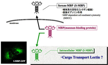
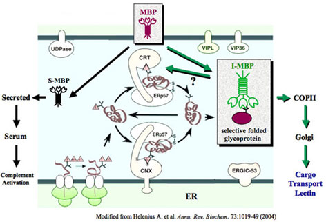
天然に存在する多くのタンパク質は、複雑な配列をもつ糖鎖によって濃密に修飾されており、近年、これらの糖鎖は核酸、アミノ酸に次ぐ第3の生命情報鎖としてその機能解析に注目があびつつある。本研究では、糖鎖に内在される生体情報の解読を目的とし、MBP(mannan binding protein)が生体内で果たす役割の解明をめざしている。MBPはマンノース、フコース、N-アセチルグルコサミンを特異的に認識するC型レクチンの一種である。MBPは一種の遺伝子にコードされ、種に肝臓で合成されるが、細胞外に分泌される血清型MBP(S-MBP)と細胞内に留まる細胞内MBP(I-MBP)が存在する。S-MBPは先天性免疫において重要な分子であるが、I-MBPの機能については未だに不明な点が多い。これまでの研究により、共焦点顕微鏡や電子顕微鏡を用いた観察から、I-MBPが小胞体(ER)の一部に特徴的な存在様式を示すことから明らかとした。さらにERとGolgiの相田を行き来する輸送小胞のタンパク質マーカーと比較、観察した結果から、I-MBPはそれら輸送小胞中に存在することが明らかとなり、糖タンパク質の細胞内輸送に関与する可能性を示した。今後、共焦点顕微鏡、電子顕微鏡を用いてさらなる詳細な観察を行い、細胞内でMBPがどの輸送小胞に局在しているかを調べ、またS-MBPとI-MBPの翻訳後修飾、分泌にどのような違いがあるかを明らかとする。次に、I-MBPと細胞内において相互作用するタンパク質の同定を行う。さらにI-MBPとの結合、解離のメカニズムなどについて表明プラズモン共鳴法を用いた詳細な解析を行うことにより、I-MBPが細胞内の環境において、新生糖タンパク質と結合、解離をすることを示す。これらの研究により、I-MBPが担う糖タンパク質の品質管理における役割が明確になると考えられる。本研究遂行には申請者が所属する研究室の持つ糖鎖生物学的研究手法だけでは十分ではなく、細胞内タンパク質の選択的輸送機構に関する専門的な細胞生物学的知識と経験が必要である。従って本研究は個々の研究分野の確立している知見や技術の融合によって達成可能な研究であり、I-MBPに関する新しい機能解明が期待される。
- Subcellular Localization of I-MBP in ER and Golgi
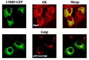
The subcellular distribution and co-localization of I-MBP with the ER and Golgi apparatus was visualized by confocal laser scan microscopy.The HuH-7 MBP-GFP stable transfectants were stained with ER marker, ERp57 and Golgi marker, p230 trans-Golgi, respectively, both followed by the corresponding secondary antibodies labeled with Alexa Fluor 568.
- Electron Microscopy Analysis for Subcelluar Localization of I-MBP
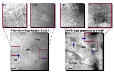
The pEGFP-N1-MBP-transfected HeLa cells were fixed and embedded.Ultrathin cryosections of the HeLa cells were stained with anti-GFP pAb, followed with 10nm colloidal immunogold labeled secondary antibody.The arrows indicate the accumlative of I-MBP in the ER, Golgi and ERGIC.
- Functional Localization of I-MBP in COPⅠ and COPⅡ Vesicles
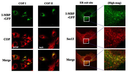
HuH-7 cells staby expressing MBP-GFP were stained with COPⅠ marker, β-COP, COPⅡ marker, Sec 31A, and ER exit site marker of COPⅡ, Sec 13, all followed by the secondary antibodies labeled with Alexa Fluor 568, and then analyzed by confocal laser scan microscopy.
- Newly Synthesized LAMP-1 Undergoes Interaction with I-MBP and BIP
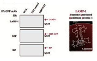
HEK293 cells were ransfected with pEGFP-N1 or pEGFP-N1-MBP vector. After 24h posttransfection, the transfectants were crosslinked with crosslinker 1 mM DSP, and the whole cell lysates(WCL) of transfectants were then immunoprecipitated by anti-GFP mAb, The immunoblots were probed with antibodies to LAMP-1, I-MBP-GFP and BIP, respectively.
- The Ca2+ Requirement for Binding of I-MBP to LAMP-1 in vivo
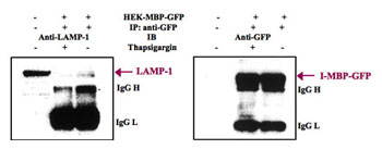
HEK293 cells transfected with pEGFP-N1 of pEGFP-N1-MBP were incubated for 12 h in the absence or presence of ER Ca+-ATPase inhibitor, 1 µM thapsigargin, and then incubated for 30 min with 1 mM DSP for crosslinking.The whole cell lysates were immunoprecipitated by anti-GFP mAb, and immunoblots were probed with antibodies to LAMP-1 and I-MBP-GFP, respectively.
- Effects of N-glycosylation Inhibitor Treatment on I-MBP / LAMP-1 Interation
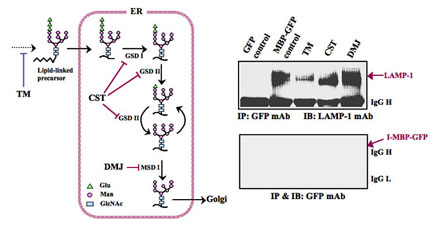
HEK293 cells transiently transfected with pEGFP-N1 or pEGFP-N1-MBP were incubated for 24 h in the absence or presence of N-glycosylation inhibitors, 2 µg/ml TM, 1 mM DMJ, respectively, and then incubated for 30 min 1 mM DSP in crosslinking buffer on a rocking platform.The immunoprecipitates with anti-GFP mAb were probed with antibodies to LAMP-1 and I-MBP-GFP, respectively.
TM, tunicamycin; CST, castanosermine; DMJ, deoxymannojirimycin;Glu, glucose; Man, mannnose; GlcNAc, N-acetylglucosamine.
- Carbohydrate-binding Assay of Wild-type and Mutant I-MBPs
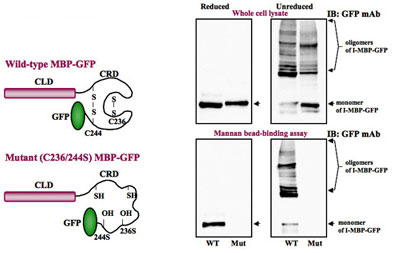
The wild-type or mutant I-MBP-GFP extracts from stable transfected HLF cells were incubated with mannan-sepharose 4B beads in the presence of 5mM CaCl2, and the bound proteins were eluted with elution buffer containing 4mM EDTA. The eluted proteins were separated by SDS-PAGE under reduced conditions, and immunoblots were probed with antibodies to I-MBP-GFP, respectively.
- Subcellular Localization of Mut I-MBP in ER but not in Golgi, COPⅠ and COPⅡ Vesicles
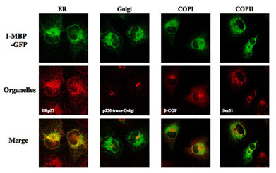
The HLF cells stably transfected with pEGFP-N1-Mutant MBP vector were stained with ER marker, ERp57, Golgi marker, p230 trans-Golgi, COPⅠ marker, β-COP and COPⅡ marker, Sec 31A, respectively, all followed by the corresponding secondary antibodies labeled with Alexa Fluor 568.
- Characterization of Biosynthesis and Differentiation of S-MBP and I-MBP in Human Hepatoma Cells
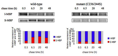
HLF cells stably expressing wild-type and mutant(C236/244S) MBP-GFPs were pulse-labeled with [35S]Met and [35S]Cys for 1 h, and were then washed and chased for up to 48 h as indicated in the figure.The medium was collected and concentrated, and the cells were solubilized with 1% NP40 lysis buffer.The immunoprecipitated S- and I-MBP-GFP with anti-GFP mAb were resolved on SDS-PAGE under reduced conditions followed by transfer to a nitrocellulose membrane, and then S-MBP and I-MBP signals of wild-type and mutant(C236/244S) on fluorograms were quantified by densitometry as shown in upper panels.The relative levels of I-MBP and S-MBP were determined comparing the expressions between I-MBP and S-MBP at te indicated chase times as shown in lower panels.
- Ultracentrifugation Analysis for Subcellular Fractions of Wild-type and Mutant I-MBPs
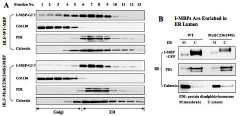
A.HLF cells stably expressing wild-type and mutant(C236/244S) MBP-GFPs were homogenized, and the post-nuclear supernatans of centrifugation onto 5-30% linear OptiPrep gradient were separated by ultracetrifugation, respectively, An aliquot of each fraction was analyzed by SDS-PAGE, and the distribution of I-MBP and organelle marker proteins was determined by direct immunoblotting.
B.ER fractions(No.6-10?) of wild-type and mutant I-MBP-GFPs were collected and incubated with 0.1 M Na2CO3, pH 11.0 to extract soluble proteins from the lumen before collecting membrances followed by centrifugation. The pallet and soluble fractions were analyzed by immunoblotting using GFP, PDI and calnexin antibodies.