|
 |
 |
|
|
Prehistory: Suprachiasmatic
nucleus is a paired cute ovoidal nuclei displaying
robust circadian oscillation |
 Okamura-lab
starts in 1995 in Kobe, when Hitoshi Okamura becomes
a professor of the Department of Anatomy II in Kobe
University School of Medicine. Thereafter, main
theme is the molecular mechanisms of mammalian circadian
clock. Before describing the achievements of the
laboratory, its prehistory is described briefly. Okamura-lab
starts in 1995 in Kobe, when Hitoshi Okamura becomes
a professor of the Department of Anatomy II in Kobe
University School of Medicine. Thereafter, main
theme is the molecular mechanisms of mammalian circadian
clock. Before describing the achievements of the
laboratory, its prehistory is described briefly.
In the Department of Anatomy II in Kyoto Prefetural
University of Medicine (Professor Yasuhiko Ibata),
Okamura encountered the dense cluster of vasoactive
intestinal peptide (VIP) producing neurons symmetrically
located just dorsal to the optic chiasma, which strongly
impressed the dignity of the suprachiasmatic nucleus
(SCN) at 1983. At that time, we analyze SCN by histochemical
and electron microscopic techniques. During these
days, we had a great effort to established two completely
original methods which will be fruitful in later
days. The first is the highly quantitative histochemistry
(Brain Research, 1987; Mol Brain Res, 1995; J. Neuroscience,
1997), and the second is the in vitro organotypic
slice culture technique for the study of the SCN
(Neuroscience, 1994: collaboration with Professor Shin-ichi
Inouye). At this time, we did not notice its
powerfulness, but both two are flowered after 10
years when mPer genes are discovered.
In France in Lyon and Gif-sur-Yvette supported by
INSERM and CNRS (1987-1989)(Professors Michel
Jouvet, Robert Naquet), we found that virtually
all SCN neurons are GABAergic.
|
1) |
Takahashi
Y, Okamura H, Yanaihara N, Hamada S, Fujita
S, Ibata Y: Vasoactive intestinal peptide immunoreactive
neurons in the rat suprachiasmatic nucleus
demonstrate diurnal variation. Brain
Res., 497: 374-377, 1989. |
2) |
Okamura H, Abitbol
M, Julien J.F, Dumas S, Berod A, Geffard M,
Kitahama K, Bobillier P, Mallet J, Wiklund
L: Neurons containing messenger RNA encoding
glutamic acid decarboxylase (GAD) in rat hypothalamus
demonstrated by in situ hybridization, with
special enphasis on cell groups in medial preoptic
area, anterior hypothalamic area and dorsomedial
hypothalamic nucleus. Neuroscience,
39: 675-699, 1990. |
3) |
Tominaga K,
Inouye S -I T, Okamura H: Organotypic slice
culture of the rat suprachiasmatic nucleus:
Sustenance of cellular architecture and circadian
rhythm. Neuroscience, 59:
1025-1042, 1994. |
4) |
Okamura H, Kawakami
F, Tamada Y, Geffard M, Nishiwaki T, Ibata
Y, Inouye S-IT: Circadian change of VIP mRNA
in the rat suprachiasmatic nucleus following
p-chlorophenylalanine (PCPA) treatment in constant
darkness. Mol. Brain Res.,
29: 358-364, 1995. |
5) |
Ban Y, Shigeyoshi
Y, Okamura H: Development of circadian VIP
rhythm in the rat suprachiasmatic nucleus. J.
Neurosci., 17, 3920-3931, 1997. |
|
|
|
Although the circadian clock genes
were identified in 1984 in Drosophila, mammalian
molecular mechanism of circadian rhythms was totally
unknown even at 1995. For example, there were many
groups believing the circadian rhythms being produced
by the networks of neurons as sleep-wake cycles, since
many molecular efforts during 13 years had failed to
isolate the clock genes in mammals. In 1997, we discovered
the human and mouse genes encoding PAS-domain (PAS,
a dimerization domain present in Per, Amt and Sim)-containing
polypeptides that are highly homologous to Drosophila period by
the collaboration of Dr. Hajime Tei in Tokyo
University (Nature, 1977). We named this gene as mPer1,
and thereafter, we proceeded the molecular dissection
solely in my laboratory, and isolated another two mammalian mPer genes
(mPer2 and mPer3) and mammalian homology
of timeless (mTim) (EMBO-J, 1998).
Subsequently, we have found that mPer1 is
light-inducible and acts as a “pendulum”, committing
the phase-shift of the circadian clock by light (Cell,
1997). Collaborating with Professor Jay Dunlap (Dartmouth
University), we found that mammalian clock is a day-type
clock similar to Neurosopra, although Drosophila’s
clock is night-type clock. Together with the discovery
of Clock gene by Professor J. Takahashi, our
studies contributed the establishment of the concept
that the mammalian circadian rhythms are generated
at the gene level.
|
1) |
Tei
H, Okamura H, Shigeyoshi Y, Fukuhara C, Ozawa
R, Hirose M, Sakaki Y: Circadian oscillation
of a mammalian homologue of the Drosophila
period gene. Nature 389,
512-516, 1997 |
2) |
Shigeyoshi Y,
Taguchi K, Yamamoto S, Takekida S, Yan L, Tei
H, Moriya T, Shibata S, Loros JL, Dunlap JC,
Okamura H: Light-induced resetting of a mammalian
circadian clock is associated with rapid induction
of the mPerl transcript. Cell 91,
1043-1053, 1997. |
3) |
Takumi T, Taguchi
K, Miyake S, Sakakida Y, Takashima N, Matsubara
C, Maebayashi Y, Okumura K, Takekida S, Yamamoto
S, Yagita K, Yan L, Young ML, Okamura H: A
light independent oscillatory gene mPer3 in
mouse SCN and OVLT. EMBO J.
17: 4753-4759, 1998. |
|
|
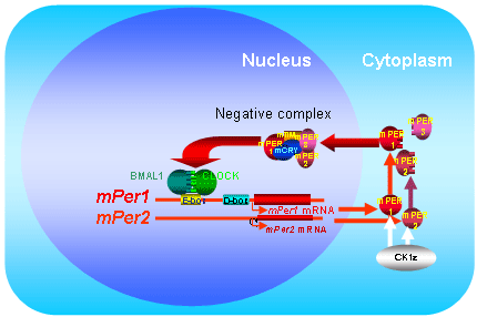
Fig. 1 Clock genes
and a model of the core feedback loop in the mammalian
cells. |
|
 |
Although we are interested in producing
knockout mice of these mPer genes to characterize
these genes, we have neither facilities to make these
animals, nor financial support at that time. So, we
were searching low cost but scientifically important
works to prove that mPer genes really have
the clock oscillating ability. It is noteworthy that
the molecular studies suggest that mammals and Drosophila
utilize similar components to generate circadian (~24
hour) rhythms. To prove that the mPer1 and mPer2 genes
indeed have the capability to generate oscillations,
we collaborated with Professor Amita Sehgal (University
of Pennsylvania) the discoverer of Drosophila timeless,
and investigated whether the introduction of mouse mPer1 and mPer2 genes
into the arrhythmic per01 mutant of Drosophila caused
functional recovery. Behavioral assays showed that
both mPer1 and mPer2 constructs driven
by Drosophila timeless promoter restored rhythms in per01 flies
that are otherwise arrhythmic due to a lack of endogenous
PERIOD protein. Thus, incorporation of mammalian mPer1 and mPer2 genes
into Drosophila mutants demonstrates that both mPer1
and mPer2 can function as clock components. This study
is unique, and indeed appreciated from some researchers,
but the recovery rate of flies are at most 20%, thus
cannot be published in leading journals at 1999 (Later
we published the results at 2002 to Genes to Cells).
Later mice homozygous for the targeted allele of mPer1 and/or mPer2 were
reported by 3 groups (Drs. Lee, Reppert and Sassone-Corsi),
showing the severely disrupted locomotor activity rhythms
during extended exposure to constant darkness. |
1) |
Shigeyoshi
Y, Meyer-Bernstein E, Yagita K, Weili F, Chen
Y, Takumi T, Schotland P, Sehgal A, Okamura
H: Restration of circadian behavioral rhythms
in a period null Drosophila mutant (per01)
by mammalian period homologues mPer1 and mPer2. Genes
Cells, 7, 163-171, 2002. |
|
|
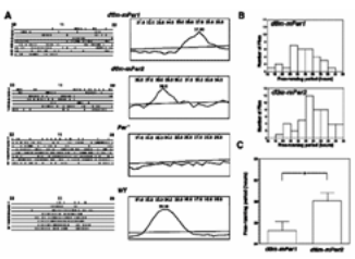
Fig. 2 Restoration
of circadian rhythm in per01 flies after the introduction
of mammalian mPer1 or mPer2 genes. A Actogram
(left) and its period length analysis
(right; period length is indicated
in hours at the top of periodigrams) of transgenic
flies (dtim-mPer1, dtim-mPer2) and controls
(per01 and wild type, WT). Circadian
time is indicated at the top of actograms, consecutive
days at left of actograms. The significance line denotes
a 0.05 level of significance. B Frequency
distribution of activity periods in tim-mPer1- and tim-mPer2-containing
transgenic flies. C Difference of
the period length in mPer1- and mPer2-
restored rhythms. From Genes Cells 7, 163-171, 2002. |
|
|
Just after the discovery of mPer genes,
the mammalian clock study goes Drosophila as
a model. In this line, mTim, mmammalian counter
part of Drosophila timeless, was soon be cloned.
Soon, clock and bmal1 are established
as positive transcription factors on mPer genes
following the negative feedback theory of Drosophila clock
machinery. However, the breakthrough comes from the
unexpected field.
Professor Gijsbertus van der Horst, a researcher
of DNA repair in Netherlands, made the knockout mice
of mammalian cryptochromes (Cry1, Cry2), and
surprisingly found that these mice were completely arrhythmic
in behavior (Nature, 1999).
Just after they got this result, we began the collaboration
whether master clock in the SCN are working or not. Okamura
and lab members trip to Rotterdam for one week experience:
fixed smples from these mice are successfully depicted.
By the quantitative in situ hybridization methods, we
found that the mPer gene oscillation was completely
abolished in the central clock (SCN) (Science, 1999).
At the same time, clock gene oscillation was abolished
in peripheral tissues (liver) of Cry deficient
mice. Then one question arises: does the peripheral clock
oscillation influence the behavioral arrhythmicity? We
performed the transplantation of wild-type SCN to arrhythmic
Cry-deficient mice, and found that the behavioral arhhythmicity
was recovered by the transplantation of wild-type SCN
(Current Biology, 2003). This rhythm emergence strongly
suggests the unnessessity of non-SCN peripheral clocks
for formation of behavioral rhythms. This result claims
the SCN as the synchronizer, but strongly supports the
SCN as the rhythm generator at behavioral rhythms, although
rhythms of peripheral clocks (endocrine, or enzyme etc)
have not been addressed. abolished it. |
1) |
Okamura
H, Miyake S, Sumi Y, Yamaguchi S, Yasui A,
Muijtjens M, Hoeijmakers JHJ, van der Horst
GTJ: Photic induction of mPer1 and mPer2 in Cry-deficient
mice lacking a biological clock. Science 286,
2531-2534, 1999. |
2) |
Sujino M, Matsumoto
K, Yamaguchi S, van der Horst G, Okamura H*,
Inouye SIT*: Suprachiasmatic nucleus grafts
restore circadian behavioral rhythms of genetically
arrhythmic mice. Current Biology 13,
664-668, 2003. (*Correspondence) |
|
|
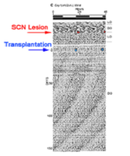
Fig. 3 Double-plotted
locomotor activity rhythms of SCN-grafted Cry1/Cry2 double
knockout mice. Arrhythmic Cry1/Cry2 double
knockout mice first lesioned SCN, then received brain
transplantation operation of neonatal SCN. Two weeks
after transplantation, activity bouts gradually consolidated
to form a free-running rhythm with a shorter than 24
hour period.
From Currrent Biology, 13, 6464-668, 2003. |
|
|
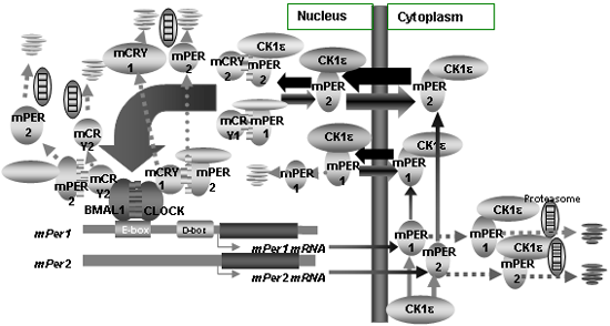
Since main components of core clock
oscillation had been revealed, a number of laboratories
began to clarify the molecular mechanisms of the mammalian
clocks in 1998-2000. The cellular circadian oscillation
in mammals to start with the transcription of two genes: mPer1 and mPer2.
Expression of these genes is stimulated by heterodimers
formed by the bHLH-PAS proteins CLOCK and BMAL1. We
demonstrated that mPER proteins made in the cytoplasm,
translocate into the nucleus (Genes Develop, 2000),
and form a negative complex comprised of mCRY1, mCRY2,
mPER1, mPER2, mPER3 and mTIM that suppresses the transcription
triggered by CLOCK and BMAL1. Importance of posttranscriptional
mechanisms including phosphorylation and degradation
are suggested. We demonstrate that mPER1 and mPER2
proteins usually shuttle between the cytoplasm and
the nucleus and are easily degraded by ubiquitination
and the proteasome pathway (EMBO-J. 2002; MCB, 2005).
Ubiquitination of mPER proteins is inhibited by the
presence of mCRY proteins. Since mCRY protein can also
be ubiquitinated when mPER proteins are absent, the
mPER/mCRY dimer is stabilized against degradation,
suppresses mPer1 and mPer2 transcription,
and shuts off mPER synthesis. Since it is speculated
that the transcription level of mPer genes
is determined essentially by the concentration of mPER/mCRY
dimer in the nucleus, re-starting mPer transcription
depends on the export of the mPER proteins out of the
nucleus by the CRM1/Exportin1 nuclear export machinery.
The consequent decrease of mPER destabilizes mCRY,
and the decrease of mCRY then allows mPer1 and mPer2 gene
transcription to restart. |
1) |
Yagita
K, Yamaguchi S, Tamanini F, van der Horst GTJ,
Hoeijmakers JHJ, Yasui A, Loros JJ, Dunlap
JC, Okamura H: Dimerization and nuclear entry
of mPER proteins in mammalian cells. Genes
Develop. 14:1353-1363, 2000. |
2) |
Yagita K, Tamanini
F, Yasuda M, Hoeijimakers JHJ, van der Horst,
GTJ, Okamura H: Nucleocytoplasmic shuttling
and mCRY dependent inhibition of ubiquitination
of the mPER2 clock protein. EMBO
J. 21, 1301ー1314, 2002. |
3) |
Yamamoto Y,
Yagita Y, Okamura H: Role of cyclic mPer2 expression
in mammalian cellular clock. Mol.
Cell Biol., 25, 1912-1921, 2005. |
|
|
Fig. 4 Phospholylation,
degradation and nuclear translocation of mPER proteins. |
|
|
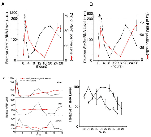
The existence of cellular clock was
indicated by the demonstration of circadian expression
of clock genes in fibroblast cell lines after the serum-shock
revealed by Schibler’s group in Geneva. Are there any
differences of autonomously rhythmic clock genes in
these cells and those in the SCN? We addressed this
question by using spontaneously immortalized mouse
embryonic fibroblasts (MEFs) from wildtype and Cry1-/-Cry2-/-
mice (Science, 2001). Both wildtype and Cry1-/-Cry2-/-
cell lines showed clock properties similar to those
found in the SCN of wildtype and Cry1-/-Cry2-/- mice,
respectively. These included: (i) temporal expression
profiles of all known clock genes, (ii) the phases
of the various mRNA rhythms (i.e. antiphase oscillation
of Bmal1 and mPer genes), (iii) the
delay between maximum mRNA levels and appearance of
nuclear mPER1 and mPER2 protein, (iv) the inability
to produce oscillations in the absence of functional mCry genes,
and (v) the control of period length by mCRY proteins.
These results strongly support the conclusion that
the components and oscillatory mechanism of central
clocks (in the SCN) and of peripheral clocks are essentially
identical. |
1) |
Yagita
K, Tamanini F, van der Horst GTJ, Okamura H:
Molecular mechanisms of the biological clock
in cultured fibroblasts. Science 292,
278-292, 2001. |
|
|
Fig. 5 Endothelin-induced
time-dependent gene expressions of mRNA (black dotted
line) and proteins (red line) in mPer1 (A)
and mPer2 (B) in mouse embryonic fibroblasts
(MEF). Note 6-8 hours difference of mRNA and proteins
in both mPer1 and mPer2. (C) (Left)
Circadian expression of Per1, dbp and Bmal1 in wild
type MEFs and mCry1-/-mCry2-/- MEFs. (Right) mCry1-/-MEF
(square) and mCry2-/-MEFs (filled circle).From Science
292, 278-292, 2001. |
|
|
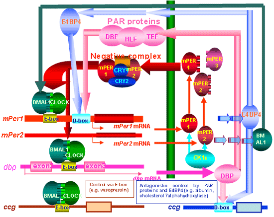
An important question is how the
time information generated by the cell clock oscillator
is transmitted to the hundreds of clock controlled
genes (ccg) that represent the output of the clock.
Two routes are proposed: the first goes directly from
the central loop of the mammalian clock to the ccgs
through E-box (CACGTG). The second route is indirect,
and regulates the antagonistic effects of PAR proteins
(DBP, HLF, TEF) and E4BP4 on the D-box (Mol. Cell Biol.,
2000; Genes Develop 2001). PAR proteins activate the
transcription of target genes by binding to the specific
sequence RTTAY GTAAY (R, purine; Y, pyrimidine) during
the day, and E4BP4 suppresses transcription of these
target genes in the night. E4BP4 and the PAR proteins
may switch back and forth in turning target gene transcription
on and off. Since many of genes contains D-box, it
is speculated over 100 genes are circadianly regulated
by D-Box. |
1) |
Yamaguchi
S, Mitsui S, Yan L, Yagita K, Miyake S, Okamura
H: Role of DBP in the circadian oscillatory
mechanism. Mol. Cell. Biol. 20:4773-4781, 2000. |
2) |
Mitsui S, Yamaguchi
S, Matsuo T, Ishida Y, Okamura H: Antagonistic
role of E4BP4 and PAR proteins in the circadian
oscillatory mechanism. Genes Develop. 15, 995-1006,
2001. |
|
|
Fig. 6 The core,
accessory and output molecular mechanisms of the mammalian
circadian clock. BMAL1/CLOCK heterodimer binds to E-box
in clock oscillating genes and ccg, and accelerates
their transcription. The core feedback loop provided
by mPer1 and mPer2 is supplemented
by an accessory PAR protein loop. DBP accelerates the mPer1 transcription,
but E4BP4 maximally expressed in counter phase to PAR
proteins suppresses mPer1 transcription. From
Cell Tiss Res 309, 47-56, 2002. |
|
|
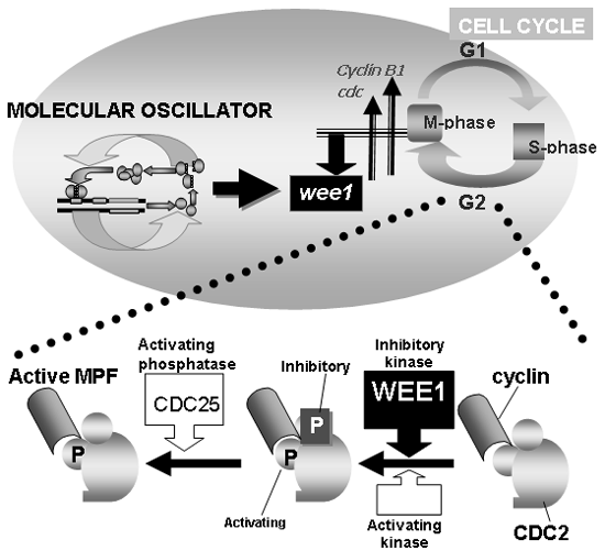
Next, we will show the examples how
core clock regulates cellular functions by analyzing
the association of circadian clock and cell cycle.
Since the life span of each cell is limited, cell growth
and mitosis are required to maintain organ or tissue
function. There is substantial evidence that circadian
rhythms affect the timing of cell divisions in
vivo: day-night variations in both the mitotic
index and DNA synthesis were found in oral mucosa,
corneal epithelium, and bone marrow. These studies
used histochemical techniques and normal physiological
conditions, so the mitotic cells comprised only a few
per cent of the cells examined. A suitable system with
a high proportion of dividing cells is required in
order to apply biochemical techniques to study the
molecular links between regeneration and the circadian
clock. We noticed that the liver provides a suitable
organ since it is known to undergo vigorous regeneration
after incomplete surgical removal. Although the cell
cycle period overall is several months in unoperated
animals, a 2/3 partial hepatectomy (PH) induces the
large majority of the remaining, pre-existing hepatocytes
to divide, and the regeneration speed is so rapid that
liver mass is restored within 7days. Moreover, expression
of mPer1 and mPer2 oscillates vigorously
in liver, and the temporal profiles and the vigorousness
of the expression of clock genes were not altered after
PH. Thus, the mouse liver is very suitable for analyzing
the molecular connection between the circadian clock
and the cell cycle.
We first compared the rate and timing of liver re-growth
after PH in mice, when PH was performed in the morning
at lights-on or in the afternoon (Science, 2003a). S-phase
kinetics, represented by the incorporation of bromodeoxyuridine
(BrdU), was similar in both morning-operated and afternoon-operated
animals. However, there was an 8 hour delay in the M-phase
(cells entering mitosis) when PH was performed at morning,
as compared to afternoon. This indicated that the timing
of the hepatectomy determines the timing of entry into
M-phase for these regenerating cells. To determine the
impact of circadian clocks on the cell cycle, clock-less
arrhythmic Cry1-/-Cry2-/- mice were subjected
to PH (Science, 1999). In these mice, mitosis was severely
impaired, and liver regeneration was severely blunted.
We performed DNA arrays and Northern blots to characterize
the molecular differences in M-phase entry and found
that cyclin B1 and cdc2 were positively
correlated, and wee1, the gene for a kinase
that inhibits mitosis by inactivating CDC2/cyclin B,
was negatively correlated to M-phase. This is interesting
since all these genes are cell cycle regulators governing
G2 to M transition, and these expression profiles correlate
well with M-phase progression. In the livers of normal
mice, there were clear circadian rhythms of wee1 expression,
with very low levels in the morning and high levels in
the night. Levels of wee1 were always high in Cry-deficient
mice, whereas levels were always low in Clock mutant
mice (Clock/Clock). There are three E-boxes
in the 5' UPR of wee1, which were, furthermore,
activated by CLOCK/BMAL1 and suppressed by PER2, PER3,
CRY1 and CRY2. These results suggest that wee1 transcription
is regulated directly by the core feedback loop through
its E-box elements. Changes in transcription of wee1 are
reflected at the protein level, influence CDC2 activity
levels, and are negatively correlated with the mitotic
peak. WEE1 phosphorylates the cyclin-dependent kinase
CDC2, a key regulator of G2 to M transition. In order
to allow entry into mitosis, CDC2 has to be dephosphorylated
by CDC25, a protein phosphatase, and it is the competition
between the activities of WEE1 and CDC25 that determines
the phosphorylation status of CDC2 (and hence the propensity
of cells to enter mitosis). Only when WEE1 levels are
low (normally in the morning) can CDC25 phosphatase successfully
antagonize the action of WEE1. These findings, taken
together, demonstrate that the circadian clock controls
the G2 to S transition via the regulation of WEE1. Together
with the report demonstrating a high incidence of lymphoma
in mice lacking the mPer2, our present study sheds light
on the association of circadian rhythms and cell division. |
1) |
Matsuo
T, Yamaguchi S, Mitsui S, Emi A, Shimoda F,
Okamura H: Control mechanism of the circadian
clock for timing of cell division.
Science 302,
255-259, 2003. (published
on line in Science Express
on 21 August 2003) |
|
|
Fig. 7 Control
of cell cycle by cell clock. Schematic representation
showing the link between the circadian clock and the
cell cycle (upper). Role of CDC2, cyclin, WEE1 and
CDC25 in making active MPF (M-phase promoting factor)
is depicted at the bottom. From J. Boil. Rhythms, 19,
388-399, 2004 |
|
|
Above studies revealed that cell
clocks govern many of the cellular functions. Then,
is the cell-time transmitted to another cells? We want
to address this issue in the SCN, since it is the sole
organ oscillate eternally. We adopt SCN-slice culture
system which we established for monitoring peptide-release
(Neuroscience, 1994). We made a SCN slice taken from
transgenic mice carrying the luciferase gene driven
by the mPer1 promoter (mPer1-luc) (Curr Biol,
2001). In this study, we discovered that the application
of NMDA, analogous to light stimuli to the living animals,
instantly altered the phase of the core clock oscillation
of slice-SCN phase-dependently. In collaborating with
Professor Masaki Kobayashi, a specialist enabling
the visualization of very weak biophoton, we succeed
to observe the rhythmic transcription of genes at the
single cell level by a cryogenic high resolution CCD
camera (Science, 2003). The SCN cells showed robust
transcription rhythms with a period length of -24 hours,
with several hundreds of cells expressing mPer1 genes
synchronously. Moreover, the individual oscillatory
cells are arranged topographically: the phase-leader
with a shorter period length is located in the dorsomedial
periventricular part of the SCN. A protein synthesis
inhibitor (cycloheximide) sets all the cell clocks
to the same phase and, following withdrawal, intrinsic
interactions among cell clocks re-establish the stable
program of gene expression across the assemblage. Tetrodotoxin,
which blocks action potentials, not only desynchronizes
the cell population, but also suppresses the level
of clock gene expression, demonstrating that neuronal
network properties dependent on action potentials play
a dominant role in both establishing cellular synchrony
and maintaining spontaneous oscillations across the
SCN. Thus, the cell-rhythm oscillation generated by
the core clock oscillatory loop is coupled and amplified
by the ordered cell-cell communications in the SCN. |
1) |
Tominaga
K, Inouye SI, Okamura H: Organotypic slice
culture of the rat suprachiasmatic nucleus:
sustenance of cellular architecture and circadian
rhythm. Neuroscience 59,
1025-1042, 1994. |
2) |
Asai M, Yamaguchi
S, Isejima H, Jounouchi M, Moriya T, Shibata
S, Kobayashi M, Okamura H: Visualization of
mPer1 transcription in vitro by luciferase
mediated bioluminescence: NMDA induces a rapid
phase-shift of mPer1 gene in cultured SCN. Current
Biology 11, 1524-1527, 2001. |
3) |
Yamaguchi S,
Isejima H, Matsuo T, Ohkura R, Yagita K, Kobayashi
K, Okamura H: Synchronization of cellular clocks
in the suprachiasmatic nucleus. Science 302,
255-259, 2003. |
|
|
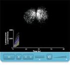
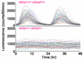
Fig. 8 Temporal
changes in bioluminescence signals from 100 SCN cells
randomly chosen in wild-type (upper figure) and mPer1-luc-bearing mCry1/mCry2 double-knockout
mice (lower figure). Note variety pattern of peaks
in cells of wild type animals, but total absence of
rhythmic cells in Cry double knockout mice.From Science
302, 255-259, 2003. |
|
|
By what route SCN regulates the peripheral
clocks? Melatonin regulates the sympathetic nervous
system by directly acting to the SCN (J. Physiol.,
2003). More importantly, sympathetic nervous system
plays the key role. In collaborating with Professor Shigenobu
Shibata, we demonstrated that hepatic gene expression
of clock genes was regulated by the sympathetic nerves
(PNAS, 2003). Moreover, we demonstrated that SCN regulates
the circadian expression of adrenal corticosterone
by the activation of various genes by the route of
innervating sympathetic adrenal nerve (Cell Metabolism,
2005). Yes, sympathetic nerve conveys the time of central
clock to peripheral organs, and the adrenal gland is
the key organ transforming circadian signals from nerve
signals to the endocrine signals. Released corticosterone
may tune the phase of peripheral clocks. |
1) |
Terazono
H, Mutoh T, Yamaguchi S, Kobayashi M, Akiyama
M, Udo R, Ohdo S, Okamura H, Shibata S: Adrenergic
regulation of clock gene expression in the
mouse liver. Proc. Natl. Acad Sci.
USA 100, 6795-6800, 2003. |
2) |
Ishida A, Mutoh
T, Ueyama T, Bando H, Masubuchi S, Nakahara
D, Tsujimoto G, Okamura H: Light activates
the adrenal gland: Timing of gene expression
and glucocorticoid release. Cell
Metabolism, 2, 297-307, 2005. |
|
|
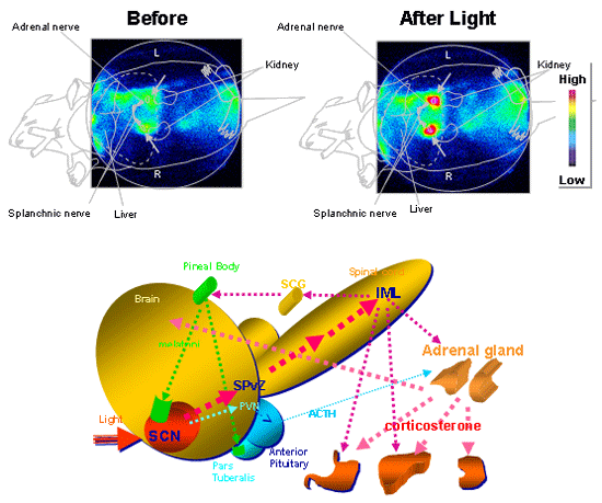
Fig. 9 (Upper
figure) Light exposure induces the high
level of expression of luciferase luminescence in
the adrenal gland (arrows) in mPer1-luc mice.
(Lower figure) The schematic representation
of the neuronal routes how light resets adrenal or
pineal hormones. IML, intermediolateral cell collumn,
PVN, paraventricular nucleus of the hypothalamus,
SCG, superior cervical ganglion, SPvZ, subparaventricular
zone.Upper figure is from Cell Metabolism, 2, 297-307,
2005. |
|
|
Monitoring bioactive markers in the
brain is fundamental to clarify the circadian time-keeping
system in the brain. In collaborating with Professor Daiichiro
Nakahara, we first devised a new method to monitor
mammalian melatonin in the cerebral ventricle by utilizing
the lintracerebral microdialysis probe. It is almost
half the century since the hormones such as melatonin
and cortisol have the diurnal rhythms. However, the
analysis of dirunal secretion of hormonesis hampered
by that there are no system to record continuously
in a single body. We developed the microdialysis system
to analyze the melatonin secretion for several months.
By this method, we have revealed for the first time
amine-dependent and nerve-independent rhythms. This
approach will became more powerful when applied tot
the genetically altered mice. |
1) |
Nakahara
D, Nakamura M, Iigo M, Okamura H: Bimodal circadian
secretion of melatonin from the pineal gland
in a living CBA mouse. Proc. Natl.
Acad. Sci. USA 100, 9584-9589,
2003. |
|
|
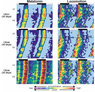
Fig. 10 A double
plot of melatonin levels and locomotor acivity for
three mice in LD and DD cycles. Note parallel change
of melatonin levels of locomotor activity in both LD
and DD conditions. Melatonin was measured from the
cerebrospinal fluid by the transverse microdialysis
probe in the pineal glands. From Proc. Natl. Acad.
Sci. USA 100, 9584-9589, 2003. |
|
|
Finally we tried to monitor the gene
expression of clock gene st real time by inserting
the optical fiber directly to the SCN in the brain
in freely moving mice. We inserted an optical fiber
just above the SCN of mPer1-luc transgenic
mice and succeeded to record for several days oscillating
luminescence that accurately mirrored native mPer1 mRNA
expression. By this method, we first revealed mPer1
gene is activated in the day time, and rest in the
night time. These data not only for the first time
demonstrate a ticking biological master clock in the
brain of a living mammal, they also show that real-time
optical imaging of gene expression is a new powerful
tool to analyze the higher nervous function of the
brain including sleep/wake cycles(Nature, 2001). |
1) |
Yamaguchi
S, Kobayashi M, Mitsui S, Ishida Y, van der
Horst GTJ, Suzuki M, Shibata S, Okamura H:
View of a mouse clock gene ticking. Nature 409,
684, 2001. |
|
|
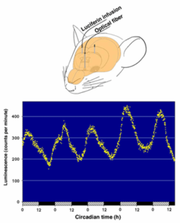
Fig. 11 (Upper
figure) Location of the input end of the
optical fiber in the mouse brain. (Lower
figure) Circadian fluctuation of luminescence
in the SCN. A mPer1-luc transgenic mouse,
previously housed under a 12h light/12h dark cycle,
was continuously infused with luciferin (10 µM
in artificial cerebrospinal fluid) at a rate of 15 µl/h.
Luminescence was recorded under constant dark conditions
via an optical fiber positioned above the SCN. One
dot represents an average of the values of 5 minutes. Hatched and closed
bars at the bottom of the figure represent subjective
day and subjective night, respectively.From Nature
409, 684, 2001, and Cell Tiss Res 309, 47-56, 2002. |
|
|
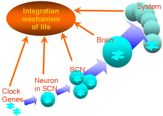
Since Human Genome Project has finished,
the revolution of medicine is now going on: now all
the history of diseases and behavior of each person
from birth to death is referenced to its genome information.
However, even this type of correspondence studies develop,
there will be no progress how genomic information is
reflected to behavioral level. The functional significance
of circadian biology exists at this point: the unique
feature of circadian biology is that the gene transcription
occurs in the SCN reflects the behavioral and physiological
rhythms almost in a perfect state. This means that
the clock gene oscillation generated by the core loop
in each SCN neuron is coupled and amplified at the
level of the SCN neuronal nuclei. From here harmonized
strongly oscillating activities are spread into the
whole brain and to all those peripheral organs, which
contain peripheral clocks. As a final result, circadian
changes are induced in behavior and hormone secretion.
Even though each neuron in the SCN generates circadian
oscillation, the system of amplification and transmission
needs to be well organized to effectively transmit
the time information to the peripheral organs. The
real time monitoring of clock gene oscillation at gene,
cell, tissue, brain, and system levels will answer
the question of how the time signal is born and integrated
to the upper layers of life. Investigations of biological
clocks open the fascinating perspective to analyze
the integrational mechanism of TIME in a vertical arrangement,
providing a bridge between single genes and the living
organism as a whole.
In May 2007, we moved from Kobe University to Kyoto University.
The new laboratory is Department of Systems Biology in
the faculty of Pharmaceutical Science. Here, we want
to reset our work to clarify “How time is generated
and tuned”. We hunt this esoteric secret poco a
poco by believing a fortune to Ride the Rhythm. |
|
Fig. 12 Integration
mechanism of rhythm. “Gene” depicts rhythmic transcription
of mPer1 and mPer2. “Cell”
represents neuronal electrical activities of single
SCN neurons. “SCN” indicates the local neuronal
and glial circuits. “System” symbolizes behavior
and hormonal secretion. |
|
|
|